

Descriptions
Product highlights
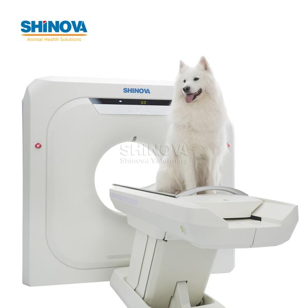
l 76cm gantry size and 65cm extended FOV enabled Luna series CT with simulation and
biopsy capabilities.
l Patent technology Vision Tec keep HD-imaging (20lp/cm@0%MTF) outperforms normal CT simulators (15 lp/cm@0%MTF)
l 32x0.625mm detector design enables true 16/32 slice acquisition provides more clarity than interpolation reconstruction
l 5.3MHU tube, 0.5s rotation speed, 250kg patient load enables a greater range of patients and applications
l Upgradable to 64-slice CT scanner, scalable based on customer requirements and budget
Specifications
1 Gantry
Aperture
70cm
One-button positioning
Preset 3 protocols
Scan speed/360°
0.7*、0.8*、0.9、1.0、1.5、2.0s
Scan FOV
43cm
Tilt range
Digital tilt±45° (step0.1)
Auto voice
Support
2.Patient Table
Max.horizontal travel range
1600mm
Horizontal scannable range
50mm~1500mm
Horizontaltravel speed
1~200mm/s
Vertical table travel range
522mm~878mm
Max.table load
206kg
One-key patient table release
Support
Patient table cradle switches
Support
3. X-ray Tube
Anode heat capacity
3. 5MHU
Equivalent anode heat capacity
9.5MHU(with iDream)
Cooling rate
735kHU/min
Focal spot size
Large: 1.2mm×1.4mm
Small: 0.7mm×0.&mm
4. Generator
Power rating
42kW
Equivalent power rating
113kW (with iDream)
kV settings
70、80、100、120、140kV
mA range (Step size)
10~350mA (1mAstep)
5. Detector
Material
Solid-state GOS
No. of Detector rows
24 rows
Max. number of slices/rotation
32 (conjugated mode)
No.of detector channels per row
768
Total No.of detector elements
18432
Min.slice thickness
0.6mm
Detector width
19.2mm
Max.data sampling rate
4800 views/360°
6. Scanning Performance
Scout scan
Supports 3 modes: AP.lateral and dual;
Scannable range 50~1500mm;
Acquisition modes
32×0.6mm
32×1.2mm
16×0.6mm
16×0.1.2mm
Min slice thickness
0.6mm
Collimation width selection
19.2mm、9.6mm
Pitch factor
0.3~1.75
(Multiple selections)
Max.continuous scan time
100s
7. Image Reconstruction
Recon FOv
50~430mm;
50~600mm (Extended).
Recon matrices
512×512、768x 768、1024×1024
Recon speed
≥20ips
Display matrix
1024×1024
8. lmage Optimization Algorithm
Metal artifact reduction
Standard
Beam hardening artifact reduction
Standard
Partial volume artifact reduction
Standard
Steaking artifact reduction
Standard
Helical scan artifact reduction
Standard
Motion artifact reduction
Standard
9. lmage Quality
Spatial resolution
≥16 lp/cm @ 0% MTF;X-Y plane
≥15 lp/cm@0% MTF;Z plane
Low-contrast resolution
2mm@0.3%@25mGy;
lmage Noise
≤0.35%(Central dose≤40 mGy)
CT HU scale
Standard: -1024HU ~+3072HU
Extended: -32768HU~+32767HU
10. Computer System
CPU
lntel Xeon 4 core,4 threads, frequency 2.9GHz,cache 8.25MB
RAM
16GB ECC
Hard disk
3TB(system disk 0.3TB+image disk 0.7TB + raw data disk 2TB)
Monitor
Size: 24 inch, LCD
Resolution: 1920×1200
Brightness: 600cd/m2
Contrast 1000:1
lmages storage
≥1,300,000 images(512×512)
External storage
DVD/CD RW, USB
Printing interface
DICOM 3.0 standard
11. Dose optimization
Dedicated pediatric protocols
Standard
Auto-mA
Standard
V-Dose check
Standard
Low dose lung screening
Standard
240° exposure
Standard
V-Beam
Standard
V-Dose report
Standard
iDream lterative reconstruction
Standard
V-Bolustracking
Standard
V-Bolus timing
Standard
Note:The image quality for the area outside the standard 430mm scan field does not meet the image quality specifications shown in the technical data sheet and image artifacts may appear, depending on the anatomy scanned.
12. Clinical Applications
Routine Applications
Lung Applications
Oncology Applications
Neurology Applications
CTA Applications
Liver Applications
Others
13. Accessories
13.1 Standard accessories

Table pad
Headrest
Headrest pad
Inferior frontal belt (standard)
Inferior frontal belt(wide)
Inferior frontal belt(narrow)
Chest and abdomen belt (standard)
Chest and abdomen belt (narrow1)
Chest and abdomen belt (narrow2)
Water phantom
System phantom
Phantom support
13.2 Optional accessories

Table extension
Knee cushion
Head and arm rest
IV poles
Flat table
Laser location lamp
14. Running Environment & Siting Requirements
14.1 Dimensions & Weight
System
Length
Width
Height
Weight
Gantry
1970mm
940 mm
1850 mm
1200 kg
Table
580 mm
2400 mm
950 mm
350 kg
Console
400 mm
630 mm
630 mm
50 kg
Distributor
810 mm
435 mm
665 mm
200 kg
14.2 Siting Requirements (Recommended)

14.3 Running Environment
Scanning room dimension
Min.area: 18m2 (5000mm x 3600mm)
Recommended room size: 30m2 (6000mm x5000mm)
Temperature&Humidity
Temperature: scanning room: 20~26°C;operating room: 18~28℃
Humidity: scanning room: 30%~70%,no condensation;
operating room: 20%~80%,no condensation
Power supply requirements
Power capacity: 70kVA
Power supply option: 3 phase 380 VAC,
voltage variation: tolerance ≤+10%
Frequency: 50 Hz or 60 Hz, tolerance ≤±1Hz
Intelligent energy saving
Insitum series CT scanners are designed to be energy saving,and have further optimized standby mode, which reduces the live operation of high-voltage control and data acquisition devices.They only keep necessary components in working state. This does not affect normal start-up efficiency, yet annual power consumption is reduced by 2815kW-h when the device is turned on for 10 hours a day, 6 days a week,which is 62.5% lower than the earlier design.
Related You might also like